Magnetic resonance imaging (MRI) is an advanced imaging technique that uses a magnetic field to create very fine detailed images of the organs and tissues within the body. Heart or cardiac MRI uses special techniques to target the heart and blood vessels. Unlike X-rays or CT scans, cardiac MRI does not expose patients to radiation.
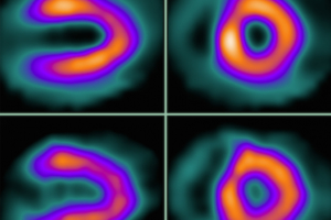
Nuclear imaging involves the use of radioactive compounds to perform diagnostic imaging examinations that can lead to the effective treatment of many diseases. Cardiac nuclear imaging refers to these diagnostic tests that are used to examine the heart. These tests are recommended for patients with unexplained chest pain, or chest pain brought on by exercise, to permit the early detection of heart disease. MHVI delivers state-of-the-art nuclear imaging technology with a comprehensive nuclear program accedited by the American Society of Nuclear Cardiology.
An echocardiogram is a diagnostic study that uses sound waves to create an image of the heart, including the heart muscle and chambers, the heart valves, the main arteries, and the pericardium (the sac around the heart). It is performed with an ultrasound wand (the transducer), which passes harmless sound waves into the body and receives them back. The images are seen on the echo machine and recorded for detailed evaluation at a later time. The test takes about 45 minutes.

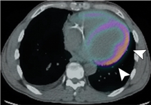
PET uses radiopharmaceuticals, or tracers, to better understand the body’s chemistry, cell function, and location of disease.


