Echocardiography
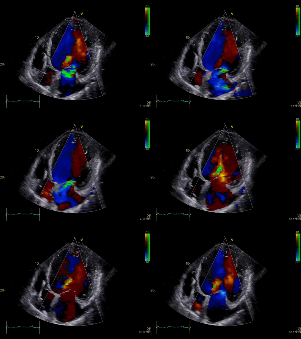
An echocardiogram is a diagnostic study that uses sound waves to create an image of the heart, including the heart muscle and chambers, the heart valves, the main arteries, and the pericardium (the sac around the heart). It is performed with an ultrasound wand (the transducer), which passes harmless sound waves into the body and receives them back. The images are seen on the echo machine and recorded for detailed evaluation at a later time. The test takes about 45 minutes.
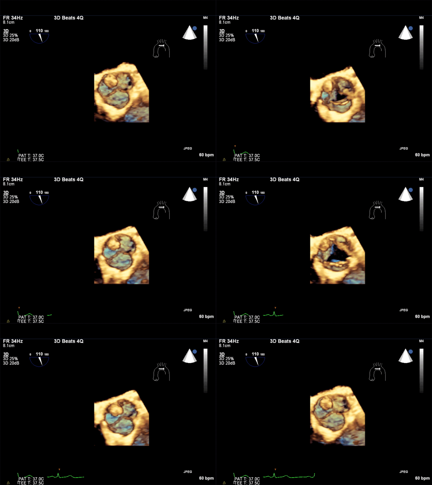
MHVI performs approximately 18,000 echocardiographic studies each year. Accredited by the Intersocietal Commission for the Accreditation of Echocardiography Laboratories (ICAEL), our full service laboratories are equipped with the latest technology in digital ultrasound imaging, providing stress echoes, transthoracic echoes, transesophageal echoes, including those with 3-D imaging and vascular ultrasounds.
How Should Patients Prepare for the Procedure?
No prepartion is required prior to the test. Patients may eat, drink, and take their usual medications prior to the test.
What Happens During the Procdure?
Patients lay on an exam table on the left side. The sonographer will place some ECG patches on the patient’s chest to record the heartbeat. The sonographer takes pictures of the heart in several places in order to obtain a complete picture. The exam will be interpreted within 24 hours, and the results will be available to the ordering physician within one to two days. At times, a saline IV may be administered to obtain additional images to check for a communication between the heart chambers. Also, an IV with a contrast agent might be placed in patients who have images that are difficult to obtain, to allow improved viewing of the heart chambers.


