Cardiac MRI
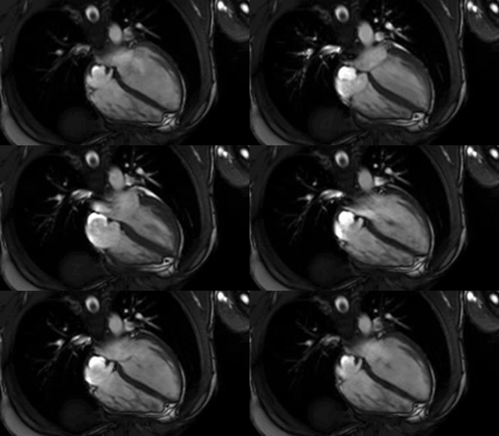
Magnetic resonance imaging (MRI) is an advanced imaging technique that uses a magnetic field to create very fine detailed images of the organs and tissues within the body. Heart or cardiac MRI uses special techniques to target the heart and blood vessels. Unlike X-rays or CT scans, cardiac MRI does not expose patients to radiation.
MHVI cardiologists and technologists obtain high-resolution images that yield excellent accuracy for the diagnosis of congenital heart defects, valvular heart disease, cardiomyopathy, heart masses, vascular disease or anomalies, as well as accurate delineation of anatomy prior to planned cardiac procedures such as coronary artery bypass surgery, pacemaker/defibrillator implantation, and catheter ablation for atrial fibrillation.
How does the procedure work?
The magnetic field from the MRI scanner temporarily aligns all the water molecules in the body. Radio waves cause these aligned particles to produce signals, which are then processed by the computer and are used to create cross-sectional MRI images. The motion imaging is very useful to study heart function and structure such as the heart walls and the heart valves. The MRI machine can combine these slices to produce 3D images that may be viewed from many different angles.
How should patients prepare for the procedure?
For a cardiac MRI study, patients typically don’t need a special preparation study, such as fasting or withholding medications, unless otherwise instructed. Once checked in, the patient changes into a gown and removes all accessories. Patients are also asked to tell the MRI technologist if there are metal or electronic devices in their bodies, which may be a potential safety hazard. If patients have anxiety or claustrophobia, they may be offered a mild sedative.
What happens during the procedure?
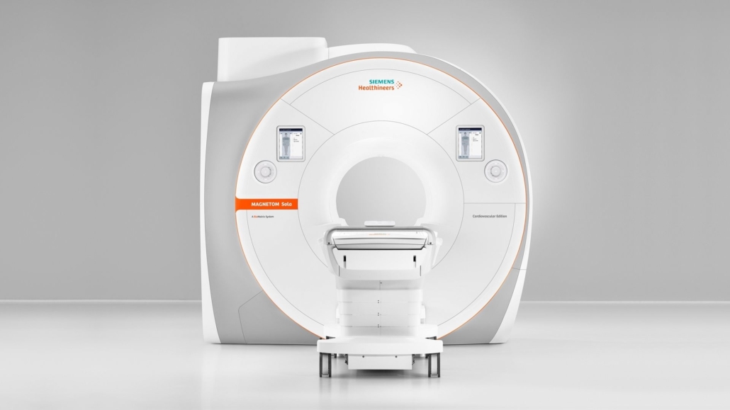
The patient lies on a table that slides into the opening of the magnetic structure. The MRI technologist monitors the patient from another room during the procedure, but can hear the patient if he or she wants to communicate by microphone. The exam itself is painless. Patients are offered earplugs or headphones to help block the noise, which can be loud. Patients do not feel the magnetic field or radio waves, and there are no moving parts to see.
What are the benefits?
Cardiac MRIs help physicians make a correct diagnosis for many heart conditions. It can very accurately diagnose an enlarged heart. For patients with a thickened heart muscle, it precisely determines the thickness of the heart muscle. A cardiac MRI can calculate how well the heart is pumping. An MRI can also accurately predict if a patient’s heart function can improve with therapy. MRI is often used in preparation for catheter ablation procedures for atrial fibrillation because it provides precise, detailed information about the anatomy.
What are the risks?
Due to the magnetic field, patients must remove metallic belongings before an MRI exam. MRI systems may damage medical devices containing metal. Patients who have these devices are generally considered unsafe for a cardiac MRI study. Patients with a history of kidney disease or kidney failure must inform the MRI technologist before undergoing an MRI. Patients who are pregnant or suspected of being pregnant should inform the technologist before undergoing an MRI.


